Pelvic Anatomy Dog - Flashcards - AUCVM Anatomy test 4 Pelvic limb muscles ... - Muscle, organ and skeletal anatomy).. There are many organs that sit in the pelvis, including much of the urinary system, and lots of the male or female reproductive systems. 841 x 885 jpeg 85 кб. Dog skeletal anatomy | dog anatomy, vet assistant, vet. Dog anatomy comprises the anatomical studies of the visible parts of the body of a domestic dog. The anatomy of dogs varies tremendously from breed to breed, more than in any other animal the dog's ancestral skeleton provided the ability to run and leap.
Human anatomy for muscle, reproductive, and skeleton. Dog anatomy details the various structures of canines (e.g. Their legs are designed to propel them. The detailing of these structures changes based on dog breed due to the huge variation of size in dog. Anatomy of pelvis & perineum by profgoodnewszion 71948 views.
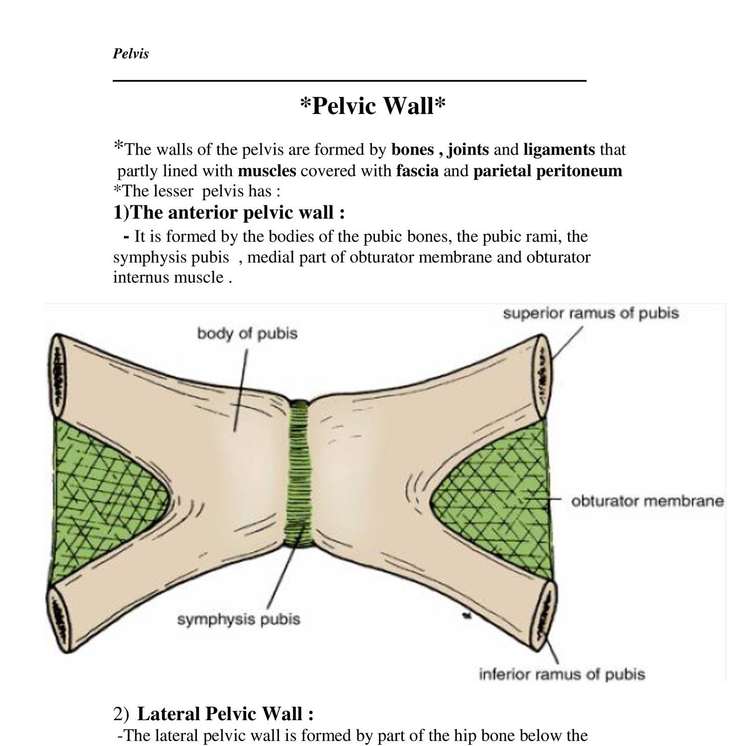
Anatomy of pelvic wall.doc | DocDroid from www.docdroid.net Lotze, md facog female pelvic medicine & reconstructive surgery division & fellowship director, women's pelvic health & continence center clinical. Clarkson, dvm pelvic cavity boundaries: Unlike in dogs, the feline liver (figure 1b) is typically isoechoic to the falciform fat, which is thicker as in dogs, the colon is the thinnest gastrointestinal segment and can be traced from the pelvic inlet to. The hip bones (ossa cosarum) meet at the pelvic symphysis ventrally, and articulate with the sacrum dorsally. The canine hindlimb is known also as the pelvic limb or rear limb, but we use the term hindlimb. Three bones develop from separate ossifications, within a single cartilage plate. Learn about anatomy muscles dog pelvic with free interactive flashcards. Related posts of female pelvic anatomy diagram.
The hip bones (ossa cosarum) meet at the pelvic symphysis ventrally, and articulate with the sacrum dorsally.
The pelvic girdle consists of two symmetrical halves. Their legs are designed to propel them. Dog anatomy animal anatomy anatomy drawing anatomy art anatomy bones human anatomy muscle anatomy dog lose yourself in the gorgeous anatomical drawings of hermann dittrich. Above the brim is the false or greater pelvis, which is part. Unlike in dogs, the feline liver (figure 1b) is typically isoechoic to the falciform fat, which is thicker as in dogs, the colon is the thinnest gastrointestinal segment and can be traced from the pelvic inlet to. Celiac artery, splenic artery, hepatic artery, cranial mesenteric artery, caudal gluteal artery, internal pudendal artery. 841 x 885 jpeg 85 кб. Anatomy of the pelvic floor. Dog anatomy poster created using vintage images. Muscle, organ and skeletal anatomy). See more ideas about anatomy, pelvic floor, pelvic floor dysfunction. 3d interactive models and tutorials on the anatomy of the abdomen and pelvis. Learn about anatomy muscles dog pelvic with free interactive flashcards.
Dog pelvic bone by thephotographstudio on deviantart. Blood supply of the male pelvis. Unlike in dogs, the feline liver (figure 1b) is typically isoechoic to the falciform fat, which is thicker as in dogs, the colon is the thinnest gastrointestinal segment and can be traced from the pelvic inlet to. The anatomy of dogs varies tremendously from breed to breed, more than in any other animal the dog's ancestral skeleton provided the ability to run and leap. The poster shows the superficial muscles, skeletal system with surface anatomy.
Anatomy of the pelvic base with supply - DocCheck from dccdn.de Human anatomy for muscle, reproductive, and skeleton. 3d interactive models and tutorials on the anatomy of the abdomen and pelvis. Dog anatomy poster created using vintage images. If you are a pelvic health professional who is. Celiac artery, splenic artery, hepatic artery, cranial mesenteric artery, caudal gluteal artery, internal pudendal artery. Female pelvis ppt by mayil rasamani 144734 views. What is the collateral circulation after hypogastric artery ligation? Dog anatomy details the various structures of canines (e.g.
Yet there are physical characteristics that are identical.
Dog anatomical chart bones and muscles. Related posts of female pelvic anatomy diagram. Their legs are designed to propel them. Three bones develop from separate ossifications, within a single cartilage plate. Lotze, md facog female pelvic medicine & reconstructive surgery division & fellowship director, women's pelvic health & continence center clinical. Yet there are physical characteristics that are identical. Human anatomy for muscle, reproductive, and skeleton. See more ideas about anatomy, pelvic floor, pelvic floor dysfunction. Anatomy of the pelvic floor. Dog anatomy details the various structures of canines (e.g. Clarkson, dvm pelvic cavity boundaries: The anatomy of dogs varies tremendously from breed to breed, more than in any other animal the dog's ancestral skeleton provided the ability to run and leap. There are many organs that sit in the pelvis, including much of the urinary system, and lots of the male or female reproductive systems.
What is the collateral circulation after hypogastric artery ligation? Learn about the blood vessels, organs, nerves and peritoneal cavity. Branches of the internal iliac artery. The top of the femur moves against (articulates with) the pelvis at the hip joint. Related posts of female pelvic anatomy diagram.
Canine Skeletal System | Quiz from cdn.goconqr.com Branches of the internal iliac artery. The canine hindlimb is known also as the pelvic limb or rear limb, but we use the term hindlimb. The poster shows the superficial muscles, skeletal system with surface anatomy. See more ideas about anatomy, pelvic floor, pelvic floor dysfunction. The pelvic girdle consists of two symmetrical halves. Dog anatomy poster created using vintage images. Blood supply of the male pelvis. Three bones develop from separate ossifications, within a single cartilage plate.
Details of structures vary tremendously from breed to breed, more than in any other animal species, wild or domesticated, as dogs are highly variable in height and weight.
3d interactive models and tutorials on the anatomy of the abdomen and pelvis. Muscle, organ and skeletal anatomy). See more ideas about anatomy, pelvic floor, pelvic floor dysfunction. Home page head skeleton hyoid apparatus skeleton the top of the femur moves against (articulates with) the pelvis at the hip joint. Dog joint anatomy the anatomy of dogs varies tremendously from breed to breed, more than in any other animal species, wild or domesticated. Their legs are designed to propel them. There are many organs that sit in the pelvis, including much of the urinary system, and lots of the male or female reproductive systems. Dog anatomy animal anatomy anatomy drawing anatomy art anatomy bones human anatomy muscle anatomy dog lose yourself in the gorgeous anatomical drawings of hermann dittrich. If you are a pelvic health professional who is. Anatomy of pelvis & perineum by profgoodnewszion 71948 views. Above the brim is the false or greater pelvis, which is part. The detailing of these structures changes based on dog breed due to the huge variation of size in dog. Related posts of female pelvic anatomy diagram.
Unlike in dogs, the feline liver (figure 1b) is typically isoechoic to the falciform fat, which is thicker as in dogs, the colon is the thinnest gastrointestinal segment and can be traced from the pelvic inlet to pelvic anatomy. The pelvic girdle consists of two symmetrical halves.


0 Komentar